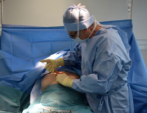
Rectal cancer surgeries require careful planning to balance complete tumor removal with preserving key bodily functions. One of the most critical considerations is whether to preserve the left colic artery, which supplies blood to parts of the colon. Preserving this artery can improve healing and reduce complications, ensuring optimal patient outcomes. Colorectal surgeon, Dr. Omar Marar, explores the impact of left colic artery preservation and its effects on long-term recovery and quality of life.
Understanding the Role of the Left Colic Artery
The left colic artery, a branch of the inferior mesenteric artery, supplies blood to the distal transverse colon, descending colon, and part of the sigmoid colon. It plays a crucial role in maintaining the vascular integrity of the colon through its connection to the marginal artery of Drummond, which provides redundancy in blood supply and minimizes the risk of ischemia. Maintaining blood flow through the left colic artery is essential for preserving colon function, especially during and after rectal cancer surgery.
Preserving the left colic artery ensures adequate blood supply to the remaining bowel after tumor resection. The risk of ischemia and anastomotic leakage significantly increases when the artery is sacrificed, as this disrupts the marginal artery’s continuity and reduces collateral circulation. Poor vascularization can delay wound healing, increase the risk of infections, and contribute to severe postoperative complications.
Patients with underlying conditions such as diabetes or atherosclerosis are particularly vulnerable to ischemic complications. Preserving the left colic artery in these patients enhances postoperative bowel function and reduces the likelihood of long-term gastrointestinal issues. Surgeons must weigh the need for oncologic clearance against the benefits of maintaining optimal blood supply to ensure the best possible outcomes.
“Achieving left colic artery preservation requires careful surgical planning and technique selection,” says Dr. Omar Marar. “We as surgeons typically choose between the high tie and low tie approaches in mesenteric artery ligation.”
The high tie approach involves ligating the inferior mesenteric artery closer to its origin near the aorta. While this facilitates lymph node dissection and improves oncologic clearance, it also increases the risk of disrupting the left colic artery’s blood supply.
The low-tie approach, in contrast, preserves the left colic artery by ligating the inferior mesenteric artery distal to its branch, maintaining blood flow to the descending colon and reducing ischemic risks.
Minimally invasive techniques such as laparoscopic and robotic surgery have significantly improved the precision of artery preservation. Laparoscopy offers enhanced visualization, allowing surgeons to avoid unintentional vascular damage, while robotic surgery provides superior dexterity and three-dimensional imaging for delicate dissections. Both approaches minimize postoperative pain, shorten hospital stays, and improve overall recovery times.
Modern intraoperative tools help surgeons assess blood flow in real time, ensuring the left colic artery remains functional. Indocyanine green (ICG) fluorescence imaging is widely used to evaluate perfusion. ICG, a dye injected intravenously, fluoresces under near-infrared light, allowing surgeons to visualize vascular integrity and confirm adequate blood supply.
Doppler ultrasound is another valuable tool for assessing blood flow velocity and confirming artery patency. These technologies help prevent ischemic complications, improve surgical precision, and enhance patient safety.
Benefits of Preserving the Left Colic Artery
Maintaining the left colic artery offers several advantages that significantly impact surgical success and recovery.
A well-vascularized anastomosis is essential for successful bowel reconnection. Preserving the left colic artery ensures a steady blood supply, reducing the risk of anastomotic leakage, which can lead to severe infections and prolonged recovery. Patients with preserved artery function typically experience better healing and fewer postoperative complications.
Ischemia can severely impair tissue health and delay recovery. By preserving the left colic artery, surgeons help maintain perfusion to critical bowel segments, lowering the risk of ischemic injury. Patients with vascular conditions benefit significantly from this approach, as their collateral circulation may already be compromised.
Sufficient blood flow supports bowel function, reducing the likelihood of postoperative irregularities. Patients who undergo surgery with left colic artery preservation experience fewer complications related to bowel motility and digestion. This contributes to improved long-term quality of life and overall patient satisfaction.
Challenges and Considerations in Left Colic Artery Preservation
Despite its benefits, left colic artery preservation presents challenges that require careful patient selection and surgical expertise.
“Not all patients are suitable candidates for left colic artery preservation,” notes Dr. Marar. Factors such as tumor location, disease stage, and vascular anatomy influence surgical decisions.”
Tumors in the lower rectum or advanced cancers may necessitate a more extensive dissection, making artery preservation less feasible. Preoperative imaging, such as CT angiography, helps assess vascular structures and guide decision-making.
The technical complexity of preserving the left colic artery demands a high level of surgical skill. Surgeons with extensive experience in rectal cancer resection and minimally invasive techniques are better equipped to perform these procedures successfully. High-volume cancer centers with advanced technology and specialized teams are more likely to achieve optimal outcomes.
Surgical complications, such as unexpected bleeding or anatomical variations, can impact artery preservation. Variations in the left colic artery’s branching pattern may complicate dissection, increasing the risk of accidental ligation. Intraoperative imaging tools, such as ICG fluorescence, help identify perfusion issues and allow for immediate surgical adjustments. Surgeons must remain adaptable to overcome challenges and ensure the best possible outcomes.
“The future of rectal cancer surgery will be shaped by advancements in precision medicine, imaging technologies, and minimally invasive techniques, further enhancing left colic artery preservation,” says Dr. Marar.
As robotic and laparoscopic surgeries continue to evolve, improved visualization and dexterity will allow surgeons to perform increasingly complex procedures with greater accuracy, minimizing vascular disruption.
Artificial intelligence and machine learning may revolutionize preoperative planning by analyzing patient-specific anatomical variations and predicting surgical outcomes, leading to more personalized approaches. Innovations in intraoperative vascular assessment, such as enhanced fluorescence imaging and real-time perfusion monitoring, will further reduce ischemic complications and improve anastomotic healing.
The integration of 3D printing and virtual reality in surgical simulations could refine training programs, equipping future surgeons with the skills necessary for meticulous artery preservation. Beyond technological progress, a shift toward multidisciplinary collaboration will refine patient selection criteria and optimize treatment strategies, ensuring the best balance between oncologic clearance and functional preservation.
As research continues to shed light on the long-term benefits of left colic artery preservation, evidence-based guidelines will evolve, standardizing surgical techniques for improved patient outcomes. With these advancements, rectal cancer surgery will become increasingly tailored, safer, and more effective, offering patients a better quality of life post-treatment.